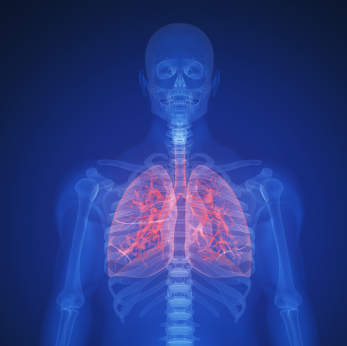
In this article, Dr Neal Navani, one of the leading practitioners offering endobronchial ultrasound in Europe, explains when and how this diagnostic procedure is used.
Contents
- What is endobronchial ultrasound?
- Why might you be advised to have endobronchial ultrasound?
- Endobronchial ultrasound and bronchoscopy
- Having endobronchial ultrasound
- Common questions that patients ask about endobronchial ultrasound
- Will I be asleep?
- Will it make me cough?
- Is endobronchial ultrasound painful?
-
How long does an endobronchial ultrasound take?
-
Can I drive home afterwards?
-
What does NICE say about endobronchial ultrasound?
-
What studies have been carried out in to endobronchial ultrasound
-
Is endobronchial ultrasound safe?
What is endobronchial ultrasound?
Endobronchial ultrasound is a relatively new diagnostic technique that can be used to investigate problems inside the lungs or in the chest area between the lungs. This is a difficult area to visualise and this used to be done using a combination of X-rays, CT scans and standard bronchoscopy. However, in order to obtain a reliable diagnosis of tuberculosis, sarcoidosis or cancer any follow-up usually had to include an invasive biopsy with an operation and an overnight stay in hospital. Today you can have a bronchoscopy with endobronchial ultrasound as a day case and you will usually be leaving for home within an hour of the procedure being completed.

Endobronchial ultrasound uses an ultrasound probe within a bronchoscope that produces images from within the airways. This has only been possible since the first miniaturised devices were developed and released in 2003. Since then the technique has been studied and subsequently used in clinical practice all over the world. It is a highly accurate and reliable outpatient test that is now accepted by the National Institute of Clinical Excellence (NICE) as an important diagnostic technique in respiratory medicine.
Why might you be advised to have endobronchial ultrasound?
Endobronchial ultrasound is used to visualise the lymph nodes in the centre of the chest (called the mediastinal lymph nodes) and to guide a needle biopsy as necessary. It is recommended if:
-
A chest CT scan has revealed that some of the lymph nodes in the centre of your chest look enlarged. This can be a sign of lung cancer, sarcoidosis or tuberculosis.
-
You have suspected tuberculosis and a chest CT scan shows enlarged lymph glands.
-
You are suspected of having sarcoidosis.
-
You have been diagnosed with lung cancer - endobronchial ultrasound is important for finding out how far your cancer has spread so that you can receive the right treatment.
Endobronchial ultrasound and bronchoscopy
An examination using endobronchial ultrasound is usually performed as part of a routine bronchoscopy. The tiny camera within the bronchoscope is fitted to the end of a thin and flexible tube, allowing the operator to look at the inside of the main airways for any areas of inflammation or damage or abnormal erosions.
The tip of the bronchoscope also contains an ultrasound transducer. This has been specifically developed for use in the lungs. When it sends out sound waves into the chest these are then reflected back to the transducer, which processes the images and sends them to a computer screen in the surgical theatre. Just as the images from the bronchoscope can be viewed in real time, the ultrasound images are also relayed instantly.

The bronchoscope tip can be moved within the airway and rotated so that the ultrasound transducer can produce the clearest view of different parts of the lung or of one of the mediastinal lymph nodes. The images can then be used to guide a needle to enable a biopsy to be taken from the area identified.
Endobronchial ultrasound combined with bronchoscopy and biopsy therefore means that it is possible to combine three different diagnostic investigations in the same procedure. The benefit of this is that the necessary diagnostic information can be obtained without the need for an operation.
Having endobronchial ultrasound
Endobronchial ultrasound involves placing the bronchoscope and ultrasound transducer into the trachea and bronchi and then using this to insert a needle into the chest if a biopsy is necessary. It is done as a day-case and you do not need to stay in hospital overnight, unless you are already being treated on a ward. You will be sitting up in bed during the test and usually able to go home 1 hour after it is completed. EBUS is very safe and is not associated with any serious complications or after effects.
Common questions that patients ask about endobronchial ultrasound
Will I be asleep?
Not usually. Patients usually have endobronchial ultrasound with sedation and with a local anaesthetic spray to completely numb the back of the throat. This avoids discomfort when the bronchoscope and tube is placed in the mouth and passed down the throat and into the airway. If you have the sedative you will still be awake but you will feel extremely relaxed and you will not remember much of the procedure afterwards. Alternatively, it is possible to carry out the procedure without sedation if the throat is thoroughly numbed.
Will it make me cough?
Most patients experience some coughing when the bronchoscope first enters the airway. This is a normal reflex but it subsides after about 20 seconds. Once the tube is inside the trachea, there are no pain sensors, meaning that patients relax as they will feel very little.
Is endobronchial ultrasound painful?
No. Some patients may have mild sore throat after the procedure but no other pain.
How long does an endobronchial ultrasound take?
The whole procedure, including the bronchoscopy, taking the ultrasound images and doing a needle biopsy can take about 45 minutes. If you have been sedated, you will then rest for a while for at least an hour and you can go home later in the day. You should always have a friend or relative with you, so that they can travel home with you and stay with you overnight to make sure that you rest until the sedative is completely out of your system.
Can I drive home afterwards?
If you have not had a sedative, yes, this is possible. Some patients cope very well without sedation and are then able to return to work once the endobronchial ultrasound is complete. However, if you have had sedation, you should not drive, operate machinery or drink alcohol for 24 hours after the procedure.
What does NICE say about endobronchial ultrasound?
NICE has published interventional procedure guidance on EBUS (guidance 254). They recommend endobronchial ultrasound as an investigative procedure and for biopsy guidance but make the point that the clinician performing the procedure should have received specific training and should have the skills to enable them to carry it out safely.
What studies have been carried out on endobronchial ultrasound?
Clinical studies have been completed in thousands of patients and the results are reassuring for patients. The results can be summarised as follows:
-
EBUS used in conjunction with a needle biopsy has a 94% sensitivity and a 100% specificity for detecting cancer within one of the chest lymph nodes. This means that it is a reliable way of diagnosing the spread of cancer to these lymph nodes.
-
Studies have also shown that EBUS plus needle biopsy has a sensitivity of 95%, a specificity of 100% and an accuracy of 89% for diagnosing the stage of lung cancer.
-
Endobronchial ultrasound and biopsy is just as reliable as surgery in detecting and staging lung cancer. It is more sensitive, more specific and more accurate than other methods, including CT scanning and PET scanning.
Is endobronchial ultrasound safe?
When done by an experienced respiratory specialist who is doing many such procedures each week, endobronchial ultrasound is very safe. Published studies report that some patients experienced a small amount of bleeding at the site where the needle for the biopsy was introduced. Theoretically, lung and throat related problems such as a sore throat afterwards, coughing up a bit of blood or lung infection could occur, but the evidence suggests that this happens very, very rarely.
A medication that reduces sensation.
Full medical glossary
The withdrawal of fluid or cells from the body by suction.
Full medical glossary
The removal of a small sample of cells or tissue so that it may be examined under a microscope. The term may also refer to the tissue sample itself.
Full medical glossary
A fluid that transports oxygen and other substances through the body, made up of blood cells suspended in a liquid.
Full medical glossary
Any of the main air pipes beyond the windpipe, or trachea, which have cartilage in their wall.
Full medical glossary
A type of endoscopy examination enabling a doctor to examine the airways through an instrument called a bronchoscope
Full medical glossary
Abnormal, uncontrolled cell division resulting in a malignant tumour that may invade surrounding tissues or spread to distant parts of the body.
Full medical glossary
The basic unit of all living organisms.
Full medical glossary
A condition that is linked to, or is a consequence of, another disease or procedure.
Full medical glossary
The abbreviation for computed tomography, a scan that generates a series of cross-sectional x-ray images
Full medical glossary
The process of determining which condition a patient may have.
Full medical glossary
Wearing away of surface tissue.
Full medical glossary
Abbreviation for Eustachian tube.
Full medical glossary
An organ with the ability to make and secrete certain fluids.
Full medical glossary
intermittent claudication
Full medical glossary
Invasion by organisms that may be harmful, for example bacteria or parasites.
Full medical glossary
The body’s response to injury.
Full medical glossary
A watery or milky bodily fluid containing lymphocytes, proteins and fats. Lymph accumulates outside the blood vessels in the intercellular spaces of the body tiisues and is collected by the vessels of the lymphatic system.
Full medical glossary
Small, rounded organs of the immune system that are distributed along the lymphatic system that filter lymph, a fluid derived from the blood, and produce antibodies and a type of white blood cells, lymphocytes.
Full medical glossary
pulmonary embolism
Full medical glossary
A rare disease of unknown cause in which there is inflammation of tissues throughout the body.
Full medical glossary
The windpipe.
Full medical glossary
An infectious disease caused by the bacterium Mycobacterium Tuberculosis.
Full medical glossary
A diagnostic method in which very high frequency sound waves are passed into the body and the reflective echoes analysed to build a picture of the internal organs – or of the foetus in the uterus.
Full medical glossary
Relating to the sense of sight (vision).
Full medical glossary
A type of electromagnetic radiation used to produce images of the body.
Full medical glossary


