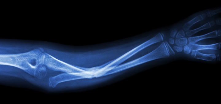
 Scientists from Trinity College Dublin have devised a revolutionary new scanning technique that produces extremely high-resolution 3D images of bones, without the need to expose patients to X-ray radiation.
The technique will have major implications for the health sector as it can be used to diagnose bone strength and provide a detailed blueprint of the extent and precise positioning of any weakness or injury. Additionally, this knowledge should help prevent the need for bone implants in many cases, and act as an early-warning system for people at a high risk of degenerative bone diseases, such as osteoporosis.
The science involves attaching luminescent compounds to tiny gold structures to form biologically safe ‘nanoagents’ that are attracted to calcium-rich surfaces, which appear when bones crack, even at a micro level. These nanoagents target and highlight the cracks formed in bones, allowing researchers to produce a complete 3D image of the damaged regions.
The research was led by the Trinity team of Professor of Chemistry, Thorri Gunnlaugsson, and Postdoctoral Researcher, Esther Surender,
Professor Gunnlaugsson said: “This work is the outcome of many years of successful collaboration between chemists from Trinity and medical and engineering experts from the Royal College of Surgeons in Ireland (RCSI). We have demonstrated that we can achieve a three-dimensional map of bone damage, showing the so-called microcracks, using non-invasive luminescence imaging. The nanoagent we have developed allows us to visualise the nature and the extent of the damage in a manner that wasn’t previously possible. This is a major step forward in our endeavour to develop targeted contrast agents for bone diagnostics for use in clinical applications.”
The work was funded by Science Foundation Ireland and by the Irish Research Council, and benefited from collaboration with scientists at RCSI, led by Professor of Anatomy, Clive Lee.
Professor Lee said: “Everyday activity loads our bones and causes microcracks to develop. These are normally repaired by a remodelling process, but, when microcracks develop faster, they can exceed the repair rate and so accumulate and weaken our bones. This occurs in athletes and leads to stress fractures. In elderly people with osteoporosis, microcracks accumulate because repair is compromised and lead to fragility fractures, most commonly in the hip, wrist and spine. Current X ray techniques can tell us about the quantity of bone present but they do not give much information about bone quality.”
He continued: “By using our new nanoagent to label microcracks and detecting them with magnetic resonance imaging (MRI), we hope to measure both bone quantity and quality and identify those at greatest risk of fracture and institute appropriate therapy. Diagnosing weak bones before they break should therefore reduce the need for operations and implants. Prevention is better than cure.”
In addition to the unprecedented resolution of this imaging technique, another major step forward lies in it not exposing X-rays to patients. X-rays emit radiation and have, in some cases, been associated with an increased risk of cancer. The red emitting gold-based nanoagents used in this alternative technique are biologically safe, indeed gold has been used safely by medics in a variety of ways in the body for some time.
Dr Esther Surender, Postdoctoral Researcher at Trinity, said: “These nanoagents have great potential for clinical application. Firstly, by using gold nanoparticles, we were able to lower the overall concentration of the agent that would have to be administered within the body, which is ideal from a clinical perspective. Secondly, by using what is called ‘two-photon excitation’ we were able to image bone structure using long wavelength excitation, which is not harmful or damaging to biological tissues.”
She added: “These nanoagents are similar to the contrast agents that are currently being utilised for MRI within the clinic, and hence have the potential to provide a novel means of medical bone diagnosis in the future. Specifically, by replacing the Europium with its sister ion Gadolinium, we can tune into the MRI activity of these nanoagents for future use alongside X-ray and computed tomography (CT) scans.”
The research is published in the journal Chem.