This article discusses an innovative radiosurgery technique, Gamma Knife®, which is used to treat primary and secondary cancers of the head and neck.This article will be of interest to anyone who has had unsuccessful surgical treatments or who may be unfit for surgery.
Contents
- What is radiosurgery?
- What is a Gamma Knife®?
- What conditions are suitable for Gamma Knife® treatment?
- Accepted indications for treatment
Introduction
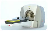
The Gamma Knife® (GK) is a machine designed to deliver radiation in a very precise manner, to treat a patient with a single dose over the course of a day rather than in a number of doses over weeks. The early prototype was first used in the late 1960s but was later developed to a model more comparable to today’s machine in the mid-1980s. As such, it is not a new invention and treatments have now stood the test of time for over 25 years. As the first radiosurgical tool available, other machines delivering single dose radiotherapy using linear accelerators (TrueBeam, CyberKnife®) take their data for the efficacy of radiosurgery from GK experience, which may, or may not be, a valid assumption given that there are differences in the homogeneity of the dose delivered by the various machines.
Although no knives are involved during treatment, GK was called a knife by its neurosurgeon inventor, Lars Leksell, to emphasise the precision with which it is able to deliver a dose of radiation to a small (or large) target in the brain. He wanted to establish the technique as an alternative to open surgery with its attendant risks, but at the same time, to maintain the management of the tumours and blood vessel anomalies suitable for radiosurgery within the same practice of neurosurgeons who were used to dealing with these patients. This is primarily the reason that GK units are normally staffed by neurosurgeons, in collaboration with a team including physicists and oncologists, whereas CyberKnife® and TrueBeam machines (using equipment able to deliver radiotherapy to any location in the body) are normally part of radiotherapy departments with treatments being prescribed by clinical oncologists.
There are currently five GK units in England (none in the rest of the UK or Republic of Ireland), approximately 35 in the rest of Europe, and at least 115 in the USA. More than 500,000 patients have benefitted from the technology since it was introduced.
What is radiosurgery?
When does radiotherapy become radiosurgery? Conventional external beam radiotherapy of the type that is delivered to most patients with malignant tumours, is given in small doses (fractions) over weeks. This is partly because the accuracy of delivery is not sufficient to concentrate the radiation on the tumour being treated without significant dosage to the surrounding tissues. By giving small doses the normal surrounding tissue has a chance to recover from the exposure to the radiation but because cancers are usually more sensitive to the effects of the radiation they are preferentially damaged, and show a greater response to treatment. The dose to the surrounding tissues, however, is sometimes associated with side effects of treatment. These effects are dependent on the type of tissue being irradiated, for example, irradiation of the bowel may lead to diarrhoea or nausea, irradiation of bone marrow to may lead to anaemia etc.
A different concept is to deliver a large dose of radiation to a tumour in a single dose as a daycase procedure (or up to 5 doses over a week – the maximum number of doses to qualify for the term radiosurgery). With more recent advances in standard radiotherapy for example intensity modulated radiotherapy or IMRT, there is a trend towards ‘hypofractionation’ (a reduction in the number of doses) but this does not equate to radiosurgery by today’s definition. By targeting a tumour in a very accurate way and delivering radiation with great precision it is possible to give a large dose, lethal to the tumour, with little risk to the surrounding normal tissues. With Gamma Knife® the treatment is normally given over the course of one day and most patients are able to return to work rapidly afterwards.
What is a Gamma Knife®?
Gamma Knife® is a trade name for the radiosurgery machine developed by Elekta in Sweden, a company set up by Lars Leksell in 1972 to develop his invention. It is suitable for tumours in the brain but can also treat areas in the head and neck below the skull base with a system referred to as ‘Extend’.
The Gamma Knife® uses Cobalt 60, a radioactive isotope, to generate the radiation used in treatment. This artificially produced isotope decays (becomes less active) over a number of years and the cobalt sources generally need to be replaced after 5–7 years as a result. Most radiotherapy machines (linear accelerators, LINACS) use electricity to generate the radiation beams needed for therapy and no radiation is generated when the machine is turned off. The Cobalt sources in the Gamma Knife® cannot be turned off, and elaborate shielding of these sources is required to prevent radiation exposure to the staff of the unit, as well as to ensure that radiation given to the patient is precisely controlled by the operator and follows the treatment plan devised. This shielding is now so good that large bunkers are no longer needed to house a Gamma Knife®, in contrast to LINACS which require thick concrete walls and a heavy metal door to prevent radiation contamination of the adjacent rooms.
What conditions are suitable for Gamma Knife® treatment?
As stated earlier, the Gamma Knife® was first developed as a way to treat lesions in the brain without the need for open surgery. This remains essentially true and most treatments are given to tumours or vascular malformations within the head. Generally tumours extending through the base of the skull and into the neck below the 2nd cervical vertebra are too low for the Gamma Knife® to treat. There is an adaptation available to allow treatments lower in the neck but generally LINAC based machines such as CyberKnife® are used in these situations.
Other limitations to treatment include the size of the tumour or malformation. Although it is possible to treat in stages for large vascular malformations, in general the larger the volume of tissue (tumour/malformation) being irradiated, the greater the risk of radiation damage to the surrounding brain. For this reason it would be unusual to offer Gamma Knife® treatments for tumours greater than 3.5cm in diameter, although the diameter may be less or slightly more than this depending on the location of the tumour and the other tumour dimensions as this affects the total volume being irradiated.
Not all tumours are suitable for treatment due to their biological properties. The majority of treatments are for well circumscribed, easily defined benign tumours, or secondary deposits of cancer within the brain. The primary brain tumours such as astrocytomas and glioblastoma multiforme are generally not suitable as these do not have a well defined ‘edge’ and tumour cells will often extend widely through the brain making the target too large, with a poor chance of controlling future growth, and a high risk of damage to the normal brain tissue that is infiltrated by the tumour.
Accepted indications for treatment:
Acoustic neuromas (vestibular schwannoma)
These are benign tumours arising from the nerves of balance (vestibular nerves) running from the inner ear to the brainstem. They are not ‘brain tumours’ as such and usually come to light as a result of hearing loss on the side of the tumour, with associated features of ‘noise’ in the ear (tinnitus) and balance disturbance. They are usually slow growing and may be present for many years before diagnosis.
There has been a major change in surgical practice over the past 15 years. Previously, most acoustic neuromas were removed surgically soon after diagnosis, but as the natural history of these tumours has become more understood, in particular their normal slow growth rate, it has been increasingly common for them to be watched for a period before any treatment is given.
Treatment, when necessary, has also changed from surgery to Gamma Knife® radiosurgery with the majority of these tumours now being treated with radiosurgery. This change is due to the results of Gamma Knife® treatment having a very high chance of arresting tumour growth coupled with a very low risk of adverse events or damage to the patient. After traditional surgery, patients often need several months off work, have facial weakness, complete loss of hearing on the side of the operation and they run the risk of infection, bleeding, stroke, leakage of cerebro-spinal fluid and rarely, death. A significant number never return to work for a variety of reasons.
GK avoids many of these risks; it is a daycase treatment and requires little or no time off work. It does not, however, remove the tumour, there may be initial tumour swelling which can be mistaken for growth before shrinkage in a significant number of cases. In addition there is a small risk of transformation of a benign tumour into a cancerous tumour from the radiation exposure. This risk is difficult to quantify with current data, but is generally estimated to be between 1 in 1000 and 1 in 10,000 cases.
Arterio-venous malformations (AVMs)
These are abnormal tangles of blood vessels that can occur anywhere in the body but cause particular problems when present in the brain. They are present from birth and may remain undiagnosed throughout life but have risks of bleeding and/or epilepsy from their presence in brain tissue. Sometimes they are found incidentally on scans of the brain performed for other unrelated problems such as ‘dizziness’ or headache.
Radiosurgery has revolutionized the treatment of AVMs, with very few now requiring surgery. The success rate of Gamma Knife® is high, with destruction of the malformation in the majority of suitable cases occurring within the first two years of treatment, although it may require 3–4 years to obliterate.
The selection of suitable cases is complex and depends on a large number of variables including the risk to the patient posed by the AVM without treatment, the size of the lesion, and the location in relation to ‘sensitive’ areas of the brain. These questions require the input of an experienced neuro-vascular team as well as the radiosurgery team, and patients should only be treated after extensive counselling about the risks posed to them from the malformation with or without treatment.
Meningiomas
These are tumours arising from the meninges (covering membranes) of the brain. They are usually benign and slowly growing but are occasionally rapidly growing and malignant. They may come to light as a result of reaching a size that raises the pressure in the head (leading to headache, failing mental capacity, visual disturbance), or from local irritative effects (epilepsy), or from damage to delicate surrounding structures (nerve deafness, double vision, balance problems etc.). Many are now identified incidentally as a result of a scan for an unrelated problem.
The decision to treat may be straight-forward in the case of a large tumour exerting significant pressure on the surrounding brain but in many cases with minor symptoms there is an option to observe the tumour with interval scanning to determine if it is growing before recommending treatment.
For those patients thought to be better with treatment than with observation, most meningiomas are still removed surgically, particularly if they are in an area that is relatively easy to operate on. The reason for this is partly to confirm the diagnosis but also because complete surgical removal of a benign meningioma almost always cures the patient, with little risk of recurrence in the future, and, in the right hands the risk of complications is low.
There are some individuals, however, who are better treated with radiosurgery. These include patients unfit for surgery due to co-existing illness or medication that makes surgery high risk. In addition, if tumours are in a very difficult area to operate on, surrounded by delicate nerves or blood vessels, complete removal is often impossible without causing unacceptable neurological problems. Furthermore, some tumours do recur after surgery, and if the recurrence is detected early while the tumour is still small, radiosurgery has a high chance of preventing additional growth.
Results from radiosurgery for meningiomas in suitable patients is good, with a greater than 90% chance of long term control of growth for benign tumours, but with less control for intermediate or malignant tumours.
Cerebral metastases
A significant number of cancer patients will suffer spread to the brain (metastatic deposits). The traditional treatment for this is to offer whole brain radiotherapy (WBRT) but increasingly, radiosurgery has been seen as an alternative for suitable individuals.
It has been estimated that perhaps 5% of individuals with cerebral secondaries would be suitable for radiosurgery but at present, the number offered this type of therapy in the UK is much lower. In the USA around 50% of the case load for a GK unit will be patients with cerebral secondaries, in the UK that number is closer to 15%. This difference in practice reflects the different views held by oncologists in Europe compared with the USA but also reflects funding difficulties for treatment within the NHS system, with many Primary Care Trusts (PCTs) refusing to fund this type of therapy despite the increasing evidence of efficacy in prolonging good quality of life for those treated.
Gamma Knife® is very unlikely to ‘cure’ any patient with cerebral secondaries but assuming that the cancer elsewhere in the body is reasonably under control and life expectancy is greater than six months, Gamma Knife® should be considered for patients with a relatively small volume of tumour metastatic to the brain (some PCTs stipulate that this must be under three deposits, but the total volume of tumour seems to be more important than the total number). For certain tumours such as breast cancer, results can be excellent in terms of prolongation of life, due partly to the newer chemotherapy agents which are able to control the cancer elsewhere in the body while GK controls the tumour in the brain. One advantage of Gamma Knife® over WBRT is that is can be repeated in the event of the development of new tumours over time.
Trigeminal Neuralgia
This is a very severe facial pain thought to be caused by compression of the trigeminal nerve (the nerve supplying feeling to the face) by an artery close to the brain stem. Most patients with this problem find that their symptoms are controlled with medication, usually carbamazepine, but if side effects are too great or the medication is not tolerated for other reasons, surgical treatment can be offered. The most effective of these techniques is surgery to move the artery away from the nerve (microvascular decompression, MVD) but a variety of other destructive treatments of the nerve exist; many of which involve an injection of the nerve ‘ganglion’, passing a needle through the cheek into an opening at the base of the skull where one of the main branches of the nerve exits. These treatments often leave the face numb as the price of pain relief but there is a high recurrence rate as the nerve later recovers.
GK is increasingly used to treat trigeminal neuralgia by targeting the trigeminal nerve and delivering a very high dose of radiation to it with great precision. It is the only radiosurgical treatment to have been assessed by the National Institute for Clinical Effectiveness (NICE) who recommended it for certain patients with neuralgia. The results are not as good as MVD but again the advantages of Gamma Knife® are that it can be used for patients unfit for surgery, is daycase and low risk. The effects of treatment take a few weeks to become apparent, usually with a gradual reduction of pain. The majority of patients will experience significant pain relief initially but there is a disappointing recurrence rate and after five years, perhaps only 50% of those treated still have good pain relief.
In the event of pain recurrence after an initial good result, retreatment with Gamma Knife® is possible, with a good chance of further pain relief but there is a greater risk of facial numbness as a result of a second dose of radiation to the nerve.
Pituitary tumours and other skull base tumours
The primary treatment for most pituitary tumours remains surgery, but the adjuvant radiotherapy treatments that have traditionally been used for controlling tumour recurrence after surgery are now being challenged by radiosurgery. In some circumstances, radiosurgery may be considered as the first line treatment, mainly in patients unfit for surgery. Good reduction of abnormally high hormone levels (acromegaly/Cushing’s disease) is usually seen after radiosurgery, but this commonly requires many months to become apparent and the effects of good surgery are better and quicker in this respect.
There are a host of less common skull base tumours which may be suitable for radiosurgery, including chordoma, chondrosarcoma and chemodectoma but these are complex decisions requiring input from a skull base surgery team in addition to Gamma Knife® clinicians.
A relatively brief review of indications cannot possibly be comprehensive. There are many rare tumours, unusual vascular lesions, and ‘functional’ problems such as epilepsy that may be suitable for Gamma Knife®. Where appropriate, a referral to the unit will be discussed in a multi-disciplinary team meeting. Self-referral is discouraged, however, as we prefer to treat in partnership with the doctors primarily involved in a patient’s care. Additionally a full medical history and copies of scans are necessary for the team to be able to make a rational decision about the role of Gamma Knife®, and these are best supplied by the referring clinicians.
An AVM or arteriovenous malformation forms when arteries connect directly to veins. This direct connection forms a high-pressure shunt or fistula. The veins are not able to handle the pressure of the blood coming directly from the arteries. The veins stretch and enlarge to create a “nidus”. Usually there are multiple feeding vessels in an AVM and many draining veins.
Full medical glossaryAn AVM or arteriovenous malformation forms when arteries connect directly to veins. This direct connection forms a high-pressure shunt or fistula. The veins are not able to handle the pressure of the blood coming directly from the arteries. The veins stretch and enlarge to create a “nidus”. Usually there are multiple feeding vessels in an AVM and many draining veins.
Full medical glossaryAssociated with the nervous system and the brain.
Full medical glossary