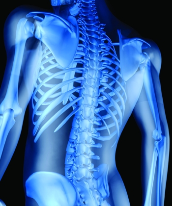
Bone health and aging
We all want to continue to look and feel well for the whole of our lives but height loss and an associated bent back (or kyphosis), is often associated with old age. However, this is not an inevitable consequence of the ageing process. The problem is most frequently caused by spine or vertebral fractures, which are surprisingly often asymptomatic. Population studies have demonstrated that fractures are present in at least 15% of European women over the age of 50. After the first vertebral fracture women (and probably men too), have a 1 in 5 chance of a second one within a year.
Effective treatments for Osteoporosis
The good news however is that osteoporosis treatment can produce a reduction in risk of up to 70%. What is more, unlike the situation with non-vertebral fractures, treatment of patients with a vertebral fracture will reduce further vertebral fracture regardless of whether or not osteoporosis is present.
DXA is quick, painless and effective
Therefore, identifying people with, and at risk of, vertebral fractures is extremely important and it is now possible using a technology called Dual Energy X-ray scanning (DXA). A DXA scan is quick and painless and more effective than normal X-rays in identifying low bone mineral density (BMD) and the corresponding risk of osteoporosis (where there is a BMD score of less than 2.5), as well as detecting pre-existing vertebral fractures using a lateral image. This process has been demonstrated to detect vertebral fractures as effectively as a lateral spine X-Ray but at a tiny fraction of the radiation dose. In fact, technically the a DXA image may often be superior to a lateral vertebral X-Ray as it avoids the problem known as “parallax” where spine bones at the outer margins of the X-ray field become distorted because of magnification and apparent overlapping of the bone margins. Lateral vertebral images are usually obtained by asking the patient to lie on their side on the DXA scanner while the scanning arm passes from the upper part of the back (thoracic spine) to the lower lumbar area, a process that takes less than 5 minutes.
Obtaining bone density measurements, combined with specialist blood tests including the bone turnover markers and information on any pre-existing spinal fractures in this way can then be used to provide appropriate and completely integrated management advice, including important lifestyle factors and, if necessary treatment options.