This article, by a Consultant Urological Surgeon, discusses the problem of kidney stones. This is an increasingly common condition and this article outlines what kidney stones are and how they can be prevented.
Contents
- What is a kidney stone?
- Who gets kidney stones?
- What causes kidney stones?
- Food and drink containing oxalate
- What are the symptoms of kidney stones?
- How are kidney stones diagnosed?
- Preventing kidney stones
Kidney stones, one of the most painful of the urologic disorders, have beset humans for centuries. Scientists have found evidence of kidney stones in a 7,000-year-old Egyptian mummy. Unfortunately, kidney stones are one of the most common disorders of the urinary tract.
What is a kidney stone?
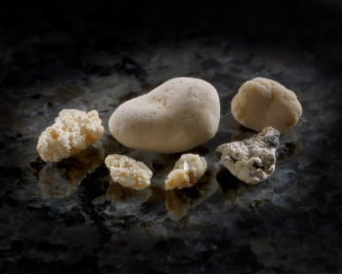
A kidney stone is a hard mass developed from crystals that separate from the urine within the urinary tract. Normally, urine contains chemicals that prevent or inhibit these crystals from forming. These inhibitors do not seem to work for everyone, so some people form stones. If the crystals remain tiny enough, they will travel through the urinary tract and pass out of the body in the urine without being noticed.
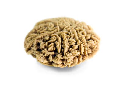
Kidney stones may contain various combinations of chemicals. The most common type of stone contains calcium in combination with either oxalate or phosphate. These chemicals are part of a person’s normal diet and make up important parts of the body, such as bones and muscles.
A less common type of stone is caused by an infection in the urinary tract. This type of stone is called a struvite infection or triple phosphate stones as they are made up of magnesium, ammonium and phosphate. Another type of stone, uric acid stones, are a bit less common, and cystine stones are rare.
Urolithiasis is the medical term used to describe stones occurring in the urinary tract. Other frequently used terms are urinary tract stone disease and nephrolithiasis. Urologists may also use terms that describe the location of the stone in the urinary tract.
Gallstones and kidney stones are not related. They form in different areas of the body. Someone with a gallstone is not necessarily more likely to develop kidney stones.
Who gets kidney stones?
For unknown reasons, the number of people in the United Kingdom with kidney stones has been increasing over the past 30 years. In the late 1970s, less than four per cent of the population had stone-forming disease. By the early 1990s, the portion of the population with the disease had increased to more than five per cent.
- Caucasians are more prone to develop kidney stones than Africans.
- Stones occur more frequently in men.
- Women most often develop kidney stones in their fifties.
The prevalence of kidney stones rises dramatically as men enter their 40s and continues to rise into their 70s. For women, the prevalence of kidney stones peaks in their 50s, however, in recent times, the peak age of stone formation in female appears to be falling. Now 13.1% of all male and 19.6% of all female stone-formers form their first stone before the age of 20 compared with 4.7% and 4.0% respectively in 1975. Once a person gets more than one stone, other stones are likely to develop.
What causes kidney stones?
Doctors do not always know what causes a stone to form. While certain foods may promote stone formation in people who are susceptible, scientists do not believe that eating any specific food causes stones to form in people who are not susceptible.
A person with a family history of kidney stones may be more likely to develop stones. Urinary tract infections, kidney disorders such as cystic kidney diseases, and certain metabolic disorders such as hyperparathyroidism are also linked to stone formation.
In addition, more than 70 per cent of people with a rare hereditary disease called renal tubular acidosis develop kidney stones.
Cystinuria and hyperoxaluria are two other rare, inherited metabolic disorders that often cause kidney stones. In cystinuria, too much of the amino acid cystine, which does not dissolve in urine, is excreted, leading to the formation of stones made of cystine. In patients with hyperoxaluria, the body produces too much oxalate, a salt. When the urine contains more oxalate than can be dissolved, the crystals settle out and form stones.
Hypercalciuria is inherited, and it may be the cause of stones in more than half of patients. In this condition, calcium is absorbed from food in excess and is lost into the urine. This high level of calcium in the urine causes crystals of calcium oxalate or calcium phosphate to form in the kidneys or elsewhere in the urinary tract.
Other causes of kidney stones are hyperuricosuria, which is a disorder of uric acid metabolism; gout; excess intake of vitamin D; urinary tract infections; and blockage of the urinary tract. Certain diuretics, commonly called water pills, and calcium-based antacids may increase the risk of forming kidney stones by increasing the amount of calcium in the urine.
Calcium oxalate stones may also form in people who have chronic inflammation of the bowel or who have had an intestinal bypass operation, or stoma surgery. As mentioned earlier, struvite stones can form in people who have had a urinary tract infection. People who take the protease inhibitor indinavir, a medicine used to treat HIV infection, may also be at an increased risk of developing kidney stones.
Food and drink containing oxalate
People prone to forming calcium oxalate stones may be asked by their doctor to limit or avoid certain foods if their urine contains an excess of oxalate.
Calcium Oxalate Stone Diet
High-oxalate foods (from highest to lowest) —
- Rhubarb, spinach, wheat germ, soybean crackers, peanuts, okra, chocolate, black Indian tea, sweet potatoes, strawberries.
Does the consumption of certain fruit juices help?
Orange juice is the most effective way to keep stones from forming. This is because it increases the alkalinity, and thus increases the potassium and citrate content of the urine which helps prevent stone formation. Grapefruit, pineapple, and apple juice also help, but not quite as much as orange juice. Cranberry and lemon juice do not increase the citrate content, although they may reduce the rate at which stones form. High levels of citrate in urine helps prevent stone formation.
What are the symptoms of kidney stones?
The first symptom of a kidney stone is extreme pain, which begins suddenly when a stone moves in the urinary tract and blocks the flow of urine.
Typically, a person feels a sharp, cramping pain in the back and side in the area of the kidney or in the lower abdomen which is called renal colic. Sometimes nausea and vomiting occur. Later, pain may spread to the groin.
If the stone is too large to pass easily, pain continues as the muscles in the wall of the narrow ureter try to squeeze the stone into the bladder. As the stone moves and the body tries to push it out, blood may appear in the urine, making the urine pink. As the stone moves down the ureter, closer to the bladder, a person may feel the need to urinate more often or feel a burning sensation during urination.
If fever and chills accompany any of these symptoms, an infection may be present. In this case, a person should contact a doctor immediately or see a urologist.
How are kidney stones diagnosed?

Sometimes “silent” stones — those that do not cause symptoms — are found on radiological imaging taken during a general health exam. If the stones are small, they will often pass out of the body un-noticed. Often, kidney stones are found on an x-ray, ultrasound or computerized tomography (CT) scan taken of someone who complains of blood in the urine or sudden pain. These diagnostic images give the doctor valuable information about the stone’s size and location. Blood and urine tests help detect any abnormal substance that might promote stone formation.
Preventing kidney stones
A person who has had more than one kidney stone may be likely to form another so, if possible, prevention is important. To help determine their cause, the doctor will order laboratory tests, including urine and blood tests. The doctor will also ask about the patient’s medical history, occupation, and eating habits. If a stone has been removed, or if the patient has passed a stone and saved it, a stone analysis by the laboratory may help the doctor in planning treatment.
The doctor may ask the patient to collect urine for 24 hours after a stone has passed or been removed. For a 24-hour urine collection, the patient is given a large container, which is to be refrigerated between trips to the bathroom. The collection is used to measure urine volume and levels of acidity, calcium, sodium, uric acid, oxalate, citrate, and creatinine—a product of muscle metabolism. The doctor will use this information to determine the cause of the stone. A second 24-hour urine collection may be needed to determine whether the prescribed treatment is working.